
App Logo

Download Our App

Shop your way

logologo
Image

How does an ectopic pregnancy occur

01/21/2026By: ICN
How does an ectopic pregnancy occur

How does ectopic pregnancy occur? (tubal pregnancy)

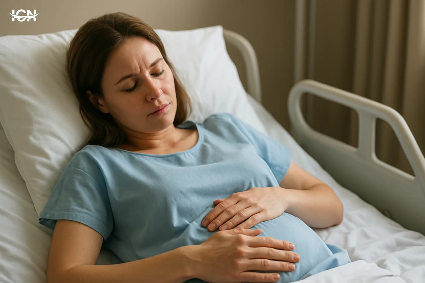
The Fallopian tube cannot accommodate the growth of the embryo, leading to the risk of a ruptured Fallopian tube and severe internal bleeding, making this a medical emergency.

What are the causes and risk factors?

Previous infections: Bacterial infections, particularly sexually transmitted infections (STIs) like Gonorrhea and Chlamydia, cause scarring, narrowing, and damage to the fallopian tubes. Statistics indicate that inflammation of the fallopian tubes significantly increases the risk. Medical history: A previous history of ectopic pregnancy, past surgeries in the pelvic or abdominal area, or hormonal disorders. Additional factors: Conditions such as endometriosis or the use of assisted reproductive technologies (IVF).

Ectopic Pregnancy Symptoms: When to Pay Attention?

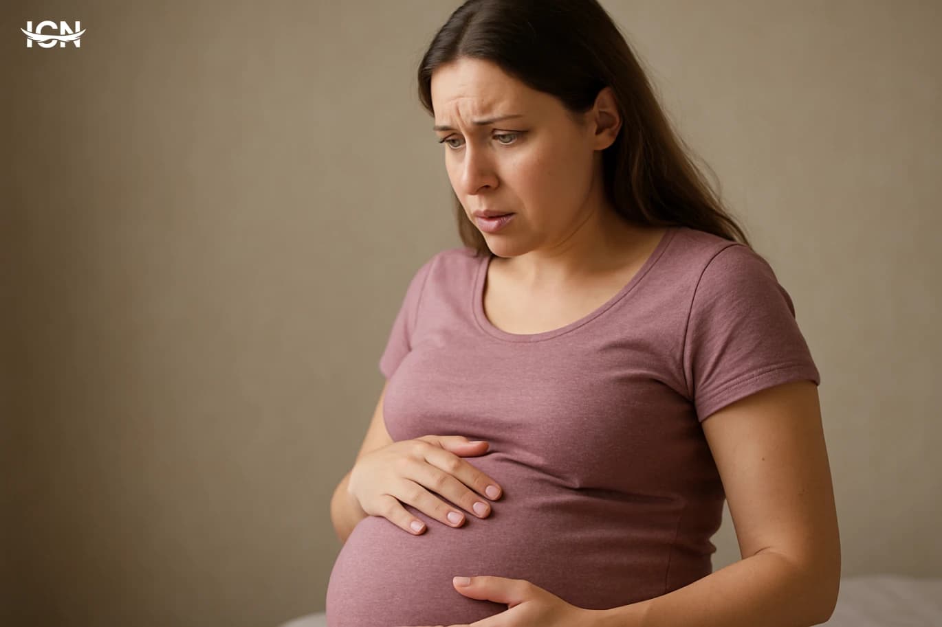
Early symptoms: Abnormal vaginal bleeding and lower abdominal pain concentrated on one side. Serious symptoms: If a tubal wall rupture occurs, the woman experiences severe and sudden pain, dizziness, paleness, and may faint due to severe internal bleeding, requiring immediate treatment.

Diagnosis and immediate treatment of ectopic pregnancy

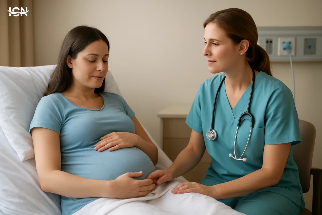
Measurement of human chorionic gonadotropin (hCG) levels in the blood. If hormone levels are low or do not double as expected, suspicion increases. Ultrasound imaging diagnosis to determine the location of the fertilized egg and ensure it is not inside the uterine cavity. The treatment for ectopic pregnancy depends on the size of the pregnancy and the hCG level. If diagnosed early and the pregnancy is small, medical treatment with methotrexate injections can be used. In advanced cases or when there is rupture of the tube, surgery (often via laparoscopy) is required to remove the pregnancy and control severe bleeding.

What is an ectopic pregnancy?

Ectopic pregnancy (extrauterine pregnancy) is defined as a failure of this journey. Instead of reaching the safe uterine cavity, the fertilized egg implants and develops outside this cavity. Doctors confirm that the vast majority of ectopic pregnancies occur within the fallopian tube, known as tubal pregnancy, accounting for 90% of recorded cases. This implantation can also occur in other rare locations, such as the ovary, cervix, or even in the abdominal cavity (peritoneal cavity rupture). This type of pregnancy poses a real danger to a woman's life, as it can never be completed.

How an Ectopic Pregnancy Occurs

This type of pregnancy can never be completed and poses a real threat to the woman's life if not quickly detected and treated. The role of the Fallopian tube in ectopic pregnancies: The Fallopian tube is the most common site for ectopic pregnancies, where fertilization occurs first. It contains fine cilia that help push the fertilized egg towards the uterine cavity. If there is damage or blockage in the Fallopian tube, the movement of the egg slows down or stops completely. As a result, the fertilized egg continues to divide and grow, attaching itself to the wall of the tube, known as a tubal pregnancy. The wall of the tube is not designed to accommodate the growth of the fetus, leading to an increase in size and the threat of the tube wall rupturing, followed by severe internal bleeding that endangers the woman's life. Causes and risk factors of ectopic pregnancy: The main reason for ectopic pregnancy is an obstacle or damage preventing the fertilized egg from passing into the uterus. Statistics show that inflammation of the Fallopian tube significantly increases the risk. Prominent risk factors leading to cilia damage and tube deformation include: Sexually transmitted infections (STIs) like chlamydia and gonorrhea, which cause chronic inflammation and scarring in the Fallopian tube. Medical history: If you have a previous history of ectopic pregnancy. Previous surgeries: Performing surgeries in the abdomen, pelvis, or on the Fallopian tube itself. Disorders of the uterine lining: Such as endometriosis, which may cause adhesions that hinder the movement of the egg. Assisted reproductive methods: In vitro fertilization (IVF) may increase the likelihood of ectopic pregnancy.

Causes and Risk Factors of Ectopic Pregnancy

Medical history and previous surgeries If you have a history of previous ectopic pregnancy, the risk of recurrence is high, with the likelihood of repeated ectopic pregnancy reaching 15%. Previous surgeries in the pelvic area or on the Fallopian tube itself may leave scars leading to obstruction or Fallopian tube damage. Previous abdominal surgeries, and even failed tubal ligations, are significant risk factors for increasing the likelihood of an ectopic pregnancy. Other risk factors In addition to causes related to the Fallopian tube, other factors that increase the likelihood of an ectopic pregnancy include: a history of endometriosis; the use of certain contraceptive methods, such as the intrauterine device (IUD), although pregnancy with it is rare; treatment through in vitro fertilization (IVF) or a history of infertility; smoking, as chemicals affect the function of the cilia responsible for moving the fertilized egg towards the uterine cavity.

Symptoms of Ectopic Pregnancy: When to Seek Medical Help?

Early warning signs that may indicate a tubal pregnancy include delayed menstruation and positive pregnancy test results. Mild vaginal bleeding or abnormal watery brown discharge. Mild to moderate pain in the lower abdomen or pelvis, often on one side of the abdomen. Slower than expected rise in Human Chorionic Gonadotrophin (hCG) levels. Emergency symptoms: Rupture of the Fallopian tube and internal bleeding If the fertilized egg continues to grow inside the Fallopian tube, the risk of tube wall rupture increases. This rupture is a medical emergency that may lead to severe and life-threatening internal bleeding, requiring immediate treatment. Symptoms of tube rupture or internal bleeding include: Sharp, sudden pain in the lower abdomen. Heavy vaginal bleeding with blood clots. Feeling dizzy and fainting due to internal blood loss. Shoulder pain, a characteristic symptom caused by nerve irritation due to collected blood in the peritoneal cavity. If you experience any of these emergency symptoms, you should immediately go to the nearest hospital to avoid complications from tube rupture.

Diagnosis of Ectopic Pregnancy: A Life-Saving Step

In normal pregnancies, the hCG hormone level nearly doubles every 48 hours. In cases of ectopic pregnancy, the hormone level rises much more slowly or may remain constant. If hCG levels are low, especially if they are less than 1500 mIU/mL, it strongly indicates the possibility of an ectopic (Extrauterine) pregnancy. Progesterone Hormone Levels are also measured, which are usually lower in ectopic pregnancies compared to normal pregnancies. 2. Ultrasound imaging for location determination Vaginal ultrasound imaging diagnosis is the primary tool to confirm the diagnosis. The doctor looks for the gestational sac within the uterine cavity. If the ultrasound shows no gestational sac in the uterus despite elevated hCG levels, it confirms the presence of an ectopic pregnancy. In many cases (more than 90%), imaging can locate the fertilized egg developing outside the uterus, often in the fallopian tube, known as a tubal pregnancy. 3. Indicator comparison: Normal vs. Ectopic Pregnancy For clarification, here is a quick comparison of the primary indicators that assist doctors in diagnosing ectopic pregnancy in the early weeks: Indicator Normal Pregnancy Ectopic Pregnancy Implantation location Intrauterine cavity Outside the uterus (often fallopian tube) hCG increase rate Doubles every 48 hours Rises slowly or remains constant Gestational sac visibility via ultrasound Seen at hCG 1500-2000 mIU/mL Not seen in the uterus despite elevated hCG levels.

Ectopic pregnancy treatment options

Data indicates that applying this early treatment significantly increases recovery rates and reduces complications, especially if the hormone level is less than 5000 IU. 2. Surgical intervention (laparoscopy) becomes a medical emergency if the ectopic pregnancy is large, if hCG levels are high, or if there are signs of imminent rupture of the canal wall. This procedure is often performed using laparoscopy, a minimally invasive surgical procedure requiring small incisions. The surgeon's goal is to remove the fertilized egg. In case of severe damage or rupture of the canal wall, the medical team may need to remove the affected tube entirely to ensure cessation of internal bleeding and prevent recurrence of ectopic pregnancy (tubal pregnancy) in the future. Immediate intervention is crucial to ensure stopping the severe internal bleeding that may result from canal rupture, which directly threatens the woman's life.

Complications of Ectopic Pregnancy: Risk of Fallopian Tube Rupture

Internal bleeding and shock as medical emergencies When a fallopian tube ruptures, there is a rapid loss of a large amount of blood in the peritoneal cavity. This severe bleeding causes a sudden and dangerous drop in blood pressure, which may lead a woman to enter a life-threatening state of shock. Therefore, any symptoms such as sudden and severe lower abdominal pain or abnormal vaginal bleeding in a woman with a missed period should be regarded as a medical emergency requiring immediate intervention. Does an ectopic pregnancy affect future pregnancies? Despite the seriousness of the condition, the success rate of early treatment reaches 80-90% in restoring health and the ability to conceive later. Many women who have experienced an ectopic pregnancy are able to achieve a normal pregnancy in the future, especially if the other fallopian tube has not suffered any damage or harm. However, it should be noted that having a history of ectopic pregnancy is one of the main risk factors for recurrence. Therefore, it is essential to closely follow up with a specialist and use ultrasound imaging and monitor hCG levels in the next pregnancy to diagnose any potential ectopic pregnancy early and prevent another tube rupture.

When should you see a doctor immediately?

Summary and Importance of Health Awareness

Ectopic pregnancy is a serious health challenge and a medical emergency requiring high awareness of its symptoms and risk factors. This condition occurs when the fertilized egg implants outside the normal uterine cavity, often up to 90% in the Fallopian tube. This type of pregnancy cannot ever be completed. A history of sexually transmitted infections or previous surgeries in the abdomen or Fallopian tube increases the likelihood of ectopic pregnancy. Early diagnosis is key to survival and is achieved by monitoring the hCG level and using ultrasound imaging to precisely determine the location of the fertilized egg implantation. Immediate treatment is essential to avoid more severe complications, such as tubal wall rupture and subsequent severe internal bleeding. Medical treatment includes Methotrexate injections, or surgical intervention via laparoscopy if the pregnancy is advanced or hormone levels are high. Remember that any delay in the menstrual cycle accompanied by abnormal vaginal bleeding or lower abdominal pain requires immediate medical attention.

Frequently Asked Questions About Ectopic Pregnancy

It may affect, especially if the treatment requires surgical intervention (such as laparoscopy) to remove the damaged fallopian tube. However, if the other tube is healthy, the chances of natural pregnancy remain high. Medication using methotrexate usually preserves the affected tube, enhancing future fertility prospects and reducing fallopian tube damage. Can ectopic pregnancy be prevented? There is no guaranteed 100% preventive method, but risk factors can be significantly reduced. Prevention focuses on avoiding main causes such as sexually transmitted infections (STIs) like chlamydia and gonorrhea, which cause infections leading to fallopian tube blockage. Also, quitting smoking and promptly treating pelvic infections is essential. What is the difference between normal pregnancy and ectopic pregnancy? The main difference lies in the implantation location of the fertilized egg. In normal pregnancy, the fertilized egg implants in the uterine cavity. In ectopic pregnancy, it implants elsewhere, often in the fallopian tube, where the embryo cannot continue to develop. What are the most important emergency signs of ectopic pregnancy? These signs require immediate treatment as they may indicate tube rupture. The main symptoms include: sudden sharp pain in the lower abdomen, severe or abnormal vaginal bleeding, feeling dizzy or faint due to internal bleeding. Diagnosis is made through ultrasound imaging and monitoring the hCG hormone level in the blood to confirm the pregnancy location.


* All articles published on this blog are sourced from various websites and are provided for informational purposes only. They should not be considered as confirmed studies or accurate information. Please verify the information independently before relying on it.

Similar ARTICLES

Why Postpartum Belly Wrinkles Persist
Many people call them “wrinkles,” but postpartum belly changes usually include a mix of skin creasing, looseness, and texture shifts. The most common contributors are stretched skin that has not fully recoiled, and stretch marks that can look like fine lines or shallow folds when the skin is relaxed. Some women notice a “crinkled” look when they sit, bend, or after a shower, while the belly may look smoother when standing. This is normal and reflects how skin behaves when it has been stretched for months. It helps to separate three issues that often get lumped together: (1) skin laxity, meaning the skin is looser and forms folds more easily; (2) stretch marks, which are small scars in the deeper skin layers and can change color over time; and (3) abdominal wall changes, such as diastasis recti, where the midline connective tissue has widened and the belly can protrude, making skin folds more visible. These can overlap, but each responds differently to time, lifestyle, and treatment. Understanding which one is dominant in your case is the first step toward realistic expectations and a practical plan.
Ninth-Month Walking Benefits
In the ninth month, many pregnant people feel torn between wanting to rest and wanting to stay active. Walking is often the most practical middle ground because it is low-impact, easy to adjust, and can be done without special equipment. In late pregnancy, the body is managing higher blood volume, increased joint laxity, and a shifting center of gravity. A steady walking routine can help maintain basic cardiovascular fitness without the strain of high-intensity workouts. What makes walking especially relevant in the final weeks is that it supports day-to-day function. Simple tasks like climbing stairs, standing for longer periods, or sleeping comfortably can become harder as the baby grows and the abdomen becomes heavier. Gentle movement helps keep the body from feeling “stuck” after long sitting, and it can reduce the sense of heaviness many people describe at the end of pregnancy. For many, a short walk after meals or in the early evening becomes a manageable habit that fits real life. Walking is not a guarantee of an easier birth, and it should never be treated as a test of endurance. Its value is in consistency and comfort. The goal in the ninth month is usually to stay mobile, support circulation, and keep stress on joints and the pelvic floor as low as possible. That is why many clinicians suggest walking as a safe option for most uncomplicated pregnancies, with individualized guidance when there are medical concerns.
Reducing Abdominal Adhesions After C-Section
Abdominal adhesions are bands of scar-like tissue that can form as the body heals after surgery. After a cesarean delivery, healing involves the uterus, abdominal wall layers, and the thin lining that covers abdominal organs. When these surfaces are inflamed or dry during surgery, or when there is bleeding and tissue handling, the body may create fibrous connections between tissues that normally slide smoothly. Many adhesions cause no symptoms and are discovered only during a later operation, but some can contribute to chronic pelvic or abdominal discomfort, pain with certain movements, or challenges during future abdominal surgery. Several factors can increase the likelihood of adhesions after a C-section. Repeat cesareans are a major contributor because each operation adds new healing surfaces and scar formation. Longer operative time, significant bleeding, infection, and emergency procedures can also raise risk. Individual healing tendencies vary as well, and conditions such as endometriosis or prior abdominal surgery may already have created adhesions. Understanding that adhesions are a common surgical outcome—rather than a rare complication—helps set realistic expectations and supports proactive steps to reduce risk where possible.
Low Belly Kicks That Tell a Story
Many pregnant people notice that kicks felt low in the belly have a distinct “pressure-and-tap” quality compared with movements higher up. This is often about anatomy and timing rather than a single meaning. Early in the second trimester, the uterus is still relatively low in the pelvis, so movements can register closer to the pubic area. As the uterus grows upward, the same baby movement may be felt in different places depending on where the baby’s back and limbs are positioned. Low sensations can also be amplified by the bladder and pelvic floor. A foot or knee pressing downward can create a sharper, more localized feeling because the pelvis is a smaller, firmer space than the upper abdomen. People who are on their feet for long periods may notice more low pressure later in the day, because gravity and fatigue can change how the uterus sits and how the baby settles. It helps to think of “low kicks” as a location report, not a diagnosis. The location can reflect baby position, the stage of pregnancy, and how your body is carrying. What matters most is the overall pattern of movement, whether it stays consistent for you, and whether any new symptoms appear alongside the change in location.
Can a Fetus Sense Light in the Womb
The uterus is not a pitch-black space, but it is far dimmer than most people imagine. Light has to pass through the abdominal wall, uterine muscle, amniotic fluid, and fetal tissues. Each layer absorbs and scatters light, so only a small fraction reaches the fetus. The amount that gets through depends on where the placenta sits, the thickness of maternal tissues, and the intensity and angle of the external light. In everyday conditions, indoor lighting is usually too weak to create a strong change for the fetus. Bright sunlight, a strong exam lamp, or a focused light source held close to the belly can produce a clearer shift. Even then, what reaches the fetus is more like a diffuse glow than a sharp beam. This matters because the fetal visual system responds primarily to changes in brightness and contrast, not to detailed images. Researchers often describe the intrauterine environment as “low light” rather than “no light.” That distinction helps explain why a fetus can show measurable responses to light late in pregnancy, while still not “seeing” the world in any meaningful way. The question is less about recognizing objects and more about whether the developing eyes and brain can register a difference between brighter and darker conditions.
Coombs Test During Pregnancy Explained
The Coombs test in pregnancy is a blood test that looks for antibodies that can attach to red blood cells. In simple terms, it helps doctors understand whether the mother’s immune system has made proteins that might react with the baby’s blood. This matters because the baby’s red blood cells can carry markers inherited from the father that are different from the mother’s. If the mother forms antibodies against those markers, the antibodies can cross the placenta and affect the baby’s red blood cells. In prenatal care, you will usually hear two related names: the indirect Coombs test and the direct Coombs test. The indirect test is the one commonly done for the pregnant person; it checks the mother’s blood for “free” antibodies circulating in the bloodstream. The direct test is typically done on the baby (or newborn) if there is concern after delivery; it checks whether antibodies are already attached to the baby’s red blood cells. Together, these tests help clinicians prevent and manage a condition called hemolytic disease of the fetus and newborn, where red blood cells break down faster than normal. Although many people associate Coombs testing only with the Rh factor, it can also detect antibodies against other blood group systems. That broader view is important because not all antibody-related issues in pregnancy are limited to Rh(D).
Labor Pain or C-Section Incision
People often compare labor pain with the pain after a cesarean, but they are not the same type of experience. Labor pain is driven by uterine contractions and cervical changes. It usually comes in waves, builds, peaks, and then eases, and its intensity can change quickly as labor progresses. It is also influenced by position, breathing, support, hydration, and pain relief options. Many describe it as intense pressure and cramping that can radiate to the back, hips, or thighs, especially during active labor. Pain after a cesarean is primarily surgical pain. It is linked to an incision through skin and abdominal layers and the healing process that follows. Instead of waves, it is more constant in the first days, and it can flare with movement such as standing up, coughing, laughing, or lifting the baby. It also comes with practical limits: getting in and out of bed, walking to the bathroom, and managing stairs. Comparing them is less useful than understanding what each involves and what recovery looks like in real life.
When the Postpartum Belly Truly Changes
Many new mothers expect the abdomen to “go back” within weeks, but postpartum changes involve several layers: the uterus shrinking, abdominal muscles recovering, skin and connective tissue tightening, and body fat redistributing. The uterus is the fastest to change; it contracts dramatically in the first days after birth and continues to shrink over the following weeks. That internal change can reduce the belly’s forward projection, yet it does not automatically restore a flat waistline. Muscles and connective tissue take longer. During pregnancy, the linea alba (the connective tissue between the rectus abdominis muscles) stretches, and the abdominal wall adapts to the growing uterus. After delivery, the tissue gradually regains tension, but the pace varies widely. Skin also behaves differently depending on genetics, age, hydration, sun exposure history, and how quickly the belly expanded during pregnancy. “Normal” is therefore not a single look or a single date. For some women, the abdomen appears close to pre-pregnancy within a few months; for others, it remains softer or more rounded for a year or longer. A realistic goal is functional recovery—comfortable movement, stable core control, and absence of pain—while appearance continues to evolve over time.
High Prolactin and Pregnancy Chances
Prolactin is a hormone produced by the pituitary gland, and its best-known role is preparing the breasts for milk production after childbirth. Outside breastfeeding, prolactin still has a normal baseline level in both women and men, and it interacts with the reproductive system through its influence on other hormones. In women who are trying to conceive, the key point is that prolactin can affect the signals between the brain, pituitary gland, and ovaries, which are responsible for regular ovulation. Prolactin levels naturally rise during pregnancy and after delivery, and they can also increase temporarily with stress, sleep changes, intense exercise, nipple stimulation, or certain medications. A single elevated reading does not always mean a chronic problem. Clinicians typically interpret prolactin results alongside symptoms, menstrual patterns, and other hormone tests. Understanding prolactin’s normal role helps explain why sustained elevation can interfere with fertility, even when other aspects of health seem stable.
Planning a Calm Hospital Birth
A hospital birth comes with clear advantages: immediate access to monitoring, anesthesia options, and specialist support if something changes quickly. At the same time, hospitals run on routines—shift changes, standard checklists, and busy units—that can make the experience feel rushed or impersonal. A practical birth plan helps you translate your preferences into simple requests that fit hospital workflows. It also reduces last‑minute decision fatigue, because you and your support person have already discussed priorities such as pain relief, mobility, and who speaks for you if you are focused on contractions. A useful plan is not a rigid script. It is a short document and a set of conversations that clarify what you want, what you want to avoid if possible, and what you are comfortable with if certain situations arise. When staff understand your baseline preferences early, they can often offer choices proactively—for example, suggesting intermittent monitoring when appropriate, or explaining when continuous monitoring is recommended. The goal is a calmer experience built on clear communication, realistic expectations, and respectful teamwork.
Planning a Calm Hospital Birth Day
Hospital birth can feel fast and unpredictable, especially when contractions intensify and decisions come quickly. A practical birth day plan is not a rigid script; it is a clear set of preferences that helps you communicate with the care team and reduces last-minute confusion. It can cover who will be with you, what information you want explained before procedures, and how you prefer comfort measures to be offered. When written in plain language and shared early, it supports smoother handoffs between nurses, midwives, and physicians across shift changes. A good plan also helps your support person know what to do. Instead of guessing, they can focus on timing contractions, offering water, reminding you to change positions, and asking questions on your behalf. Many hospitals have standard protocols, but there is still room for choice in lighting, movement, monitoring options, and newborn care routines. The goal is to align expectations, not to control every outcome. Even if the plan changes, having it makes it easier to adapt because you have already discussed priorities.
Hidden Triggers of Breathlessness in Pregnancy
Breathlessness is common in pregnancy, but it can still feel unexpected, especially when it shows up during simple tasks like climbing a few steps or talking for a long time. Part of the reason is that your body changes quickly: blood volume rises, the heart works harder, and breathing patterns shift to meet higher oxygen needs. Many people notice it earlier than they expected, sometimes in the first trimester, even before the belly is large. What often goes unnoticed is that “normal” pregnancy breathlessness can be amplified by small, fixable factors. A day of poor sleep, a stuffy room, dehydration, or a heavy meal can make breathing feel tighter without any major medical issue. The sensation can also vary by time of day, posture, and activity level, which makes it hard to pinpoint a single cause. It helps to track when it happens and what was going on beforehand: after eating, after a long car ride, in warm weather, or when lying flat. That pattern can reveal hidden triggers and guide practical changes. At the same time, any breathlessness that is new, rapidly worsening, or paired with concerning symptoms should be discussed with a clinician promptly, because pregnancy also changes how some conditions present.
Normal Pregnancy Changes That Seem Scary
Pregnancy is a planned, whole-body shift driven by hormones, increased blood volume, and the growing uterus. Many sensations feel sudden because changes happen week by week, not gradually over years. Estrogen and progesterone affect the skin, digestion, mood, and sleep. Blood volume rises significantly to support the placenta, which can change heart rate, breathing, and temperature. The uterus expands and changes posture and muscle balance, which can create new aches even if nothing is “wrong.” A useful way to reduce worry is to connect a symptom to a normal mechanism. For example, feeling warmer can be linked to higher metabolic demand and increased circulation. Feeling more tired can reflect the body building placenta tissue and adjusting to new hormone levels, especially in the first trimester. When you understand that pregnancy is a coordinated set of adaptations, many “strange” signs become easier to interpret as expected, not alarming.
By clicking the SUBSCRIBE button, you are agreeing to our Privacy & Cookie Policy If you want to unsubsribe the marketing email, please proceed to our privacy center.
© 2005-2026 ICN. All Rights Reserved.